Review by L Flood
Middlesborough, UK
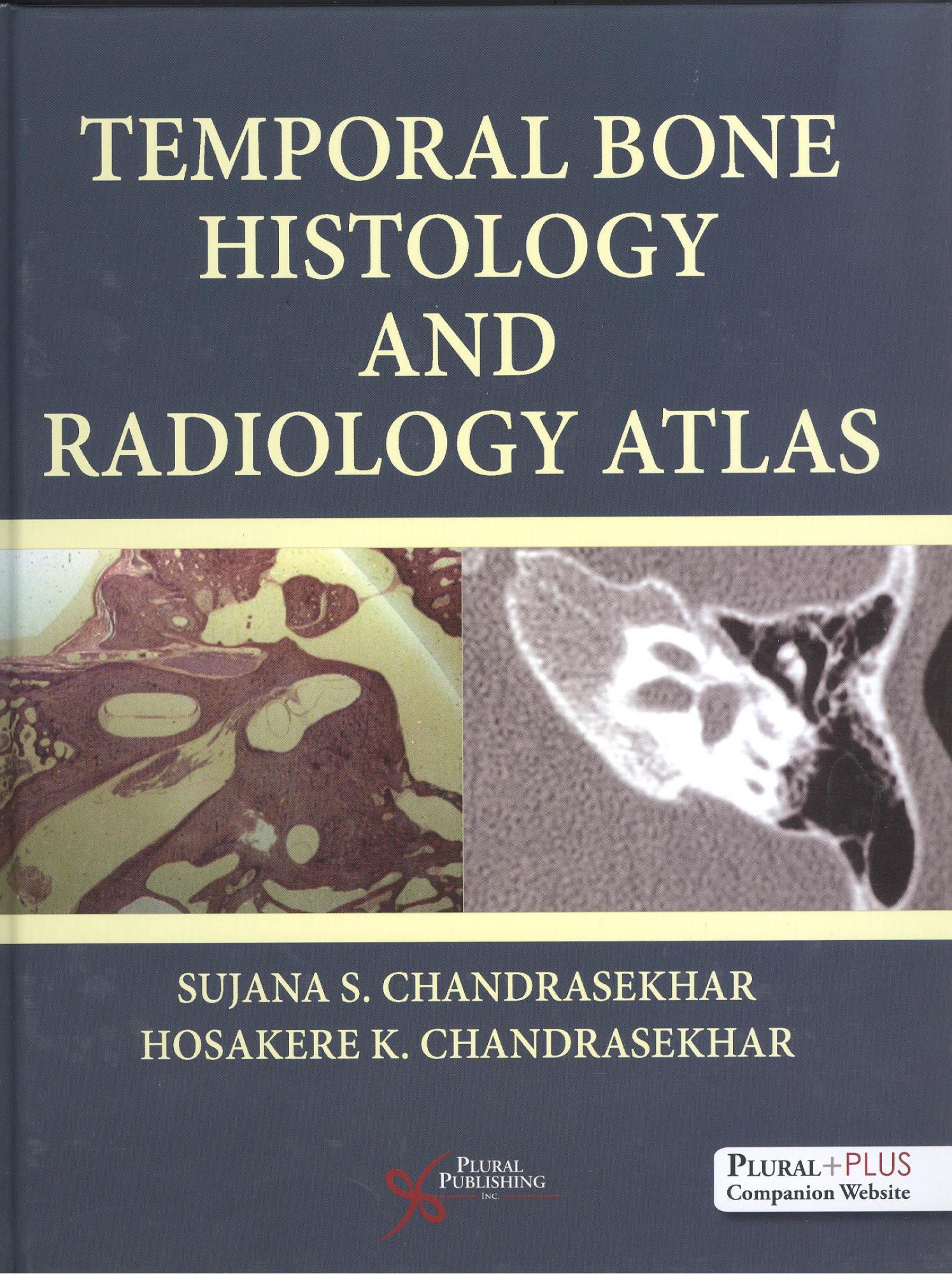
I have long been aware of the Chandrasekhar Limit, a critical mass that determines the ultimate fate of a dying star, but these are a father and daughter who have co-edited a book describing what I had thought of as largely a lost art. Those of us who trained in the late 1970s will recall the enthusiasm for establishing temporal bone collections. The 28 laboratories in the USA in 1976 are now reduced to 3, because of a lack of funding and the specialist skills required. Established in 2006, the Otopathology Research Collaboration Network unites three member laboratories in Massachusetts, California and Minnesota to continue this work. The book is based on the authors’ experience of over two decades of running an instructional course at Academy meetings and for residents at the New York University Medical School.
This is indeed an atlas, and largely concerns normal anatomy rather than disease processes. This is basic science at its best, much lamented as lacking in UK undergraduate (indeed increasingly in post-graduate) education and training.
The opening chapter is highly specialised, describing techniques for harvesting cadaver temporal bones. This is followed by a very detailed review of the preparation and analysis for histological study. The result is many a haematoxylin and eosin stained illustration, but also then a masterful review of the ultrastructure of the cochlea.
Chapter 3 suddenly leaves the ‘dead ear’ to illustrate temporal bone anatomy radiologically, in vivo (and in health, not disease, of course). Very high resolution computed tomography and magnetic resonance imaging scans are nicely printed and selected. Curiously, we are then back to osteology of the temporal bone, a subject I would have expected to be placed earlier. We now get boxed ‘clinical caveats’, pearls of clinical wisdom that are well thought out. In Chapter 5, on facial nerve anatomy, these are particularly useful, complementing the nice radiological and sectional images of anatomy. I thought a line diagram of the insane complexity of the facial nerve seemed in a very dated style, however accurate it surely is. I now note that it is taken from 1918!
The atlas idea now takes over, with far less text, but instead full page colour illustrations of sectional anatomy and the corresponding radiology shown opposite. Coronal images we are all surely familiar with, for cholesteatoma surgery. Axial sections we routinely review for deeper temporal bone disease. Sagittal images only became possible with advances in processing and are still a novelty to this reviewer. Curiously, of course, this is just the view obtained by the enthusiast wielding a drill and cutting burr!
Pathological processes are few in number and are largely concentrated in the final chapter. There is many a haematoxylin and eosin stained otosclerotic focus, and there has to be a superior semicircular canal dehiscence in any text these days. This book is encouraging in showing that this fundamental aid to studying the ear is not an anachronism, as feared. It will be of value to students of radiology and otology, and comes with individual access to supplementary materials in an online website.
Amazon Link: Temporal Bone Histology and Radiology Atlas
By purchasing books via this link you will help to fund the JLO

