Review by L Flood
Middlesborough, UK
This a topic dear to my heart, the end result being my redundant collection of over three-thousand slide transparencies, a few hundred of which I did manage to digitise, and a collection of 35 mm film cameras that do not even have scrap value now. The opening chapter describes how such images are now captured in the digital age. It does carry a nice illustration of ‘a set up used in past years’, exactly the kit I once used. Fortunately, most of us do now have access to the modern visual aids illustrated in the following Figure 1.7.
A nice Preface tells us that ‘Otoscopy alone can establish the diagnosis in some cases, parameters such as history and audiological and neuroradiological evaluation are required in others’. Otoscopy is a bit like oral surgery or even dermatology, where pattern recognition and decades of experience can make all the difference to correct and instant diagnosis. The authors do advise of course that what you see, the red blush, the white bulge, the fluid-filled ear, is ‘the tip of the iceberg’.
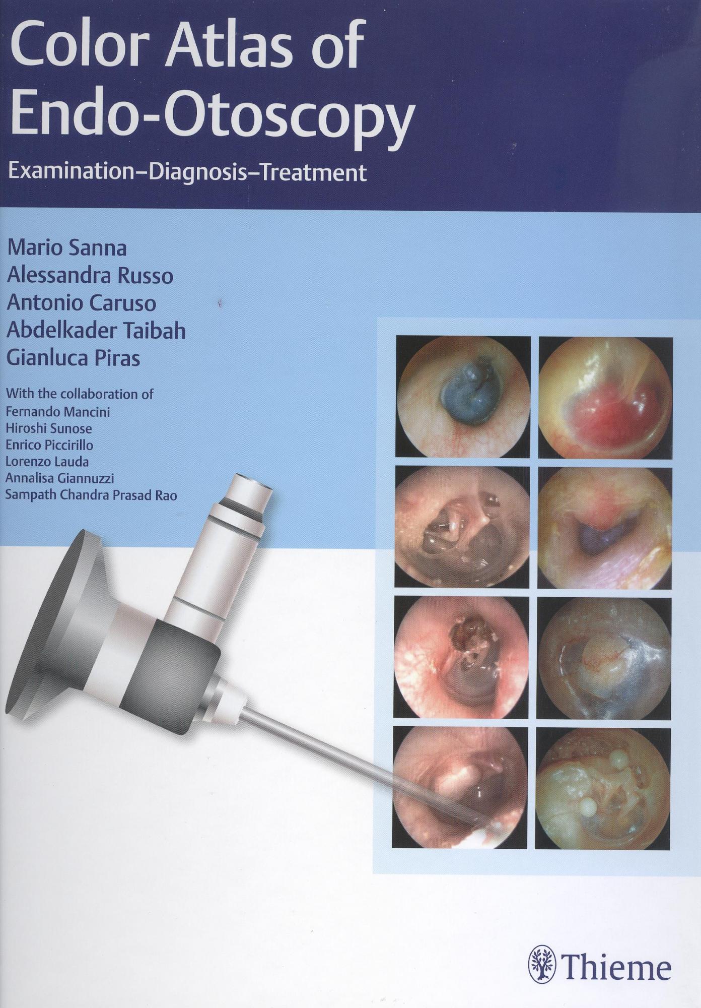
This book is far more comprehensive than the title, or even the cover, suggests. I expected to flick through countless pictures of typical tympanic membrane and middle-ear lesions, and, with over a thousand illustrations, there are plenty of those. There are countless computed tomography scans, nicely printed and clearly labelled; there are operative images, taken through the microscope, that are of superb quality. There is surprisingly detailed text on the underlying pathology, staging and even management of the various disease processes covered. A typical example is surgery of external canal exostoses, and I was relieved to read that the authors share my success rate in surgical management of post-inflammatory canal stenosis. They advise against it! I was struck by the coverage of external canal carcinoma, offering differential diagnosis, staging and detailed diagrams of tumour extension, in what is called simply an atlas.
The blue drum of cholesterol granuloma is not easily captured, but nicely shown here. Indeed, middle-ear effusion is illustrated, but with far more coverage of the countless weird and wonderful skull base tumours that may be responsible. Again, one expects nice views of ossicular disorders through perforations or retractions, of cholesteatomas and of middle-ear masses. The surprise is the coverage of tympanoplasty techniques and the excellent microscopy images of mastoidectomy (Figures 8.73–8.111). Chapter 12, ‘Rare Retrotympanic Masses’, reports precisely such, the really obscure, probably reflecting the group’s experience over 30 years, of 32 000 operations and 300 000 consultations! The final chapter ‘Postsurgical Conditions’ shows an unconvincing Schwartz sign, which I will forgive as challenging to capture, with a series of failed tympanoplasties and extruding prostheses.
I had expected to see more coverage of otoendoscopic surgery of the ear, so increasingly popular amongst the younger surgeons. Instead, this book is very much geared towards the diagnostic uses of otoendoscopy. Overall, this book is invaluable to trainees, from the youngest medical students to the over sixties, who still have much to learn, I find.
Amazon Link: Color Atlas of Endo-Otoscopy: Examination-Diagnosis-Treatment
By purchasing books via this link you will help to fund the JLO

